
Skin Cancer Treatment in Youngstown
A Frightening Fact about Skin Cancer
The incidence of skin cancer is rapidly increasing throughout the world. More than 1.3 million new cases of skin cancer are diagnosed each year in the United States alone. There are a number of factors contributing to this rise, ranging from increased time spent in the sun to the popularity of immunosupressive treatments for other diseases.
The cancers shown in the following examples are dangerously large and were selected to demonstrate the tumors’ characteristics. Many of them should have been removed much sooner. If you are concerned about a lesion on you, a relative or friend, don’t wait. Suggest they see a dermatologist today.
Types of Skin Cancer
The Malignant Melanoma:
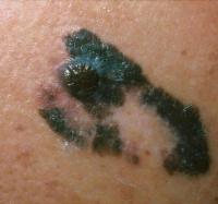 The malignant melanoma is a very serious and aggressive form of skin cancer. It is responsible for approximately eight thousand deaths a year. To learn more about malignant melanomas and see examples, click here.
The Amelanotic Malignant Melanoma:
 A particularly dangerous variant of malignant melanoma is amelanotic melanoma (“amelanotic” means without color). As the name implies, these melanomas do not have the typical dark color of a melanoma, and are therefore often overlooked. Unfortunately, they are just as serious, and need to be identified early. To learn more about amelanotic melanomas and see examples, click here.
Regression of Melanomas:
 In some cases, dark-colored melanomas may lighten in color or appear to lessen in extent through a process called regression. Just as with amelanotic melanomas, this does not lessen their severity or mean that they are less of a threat to your health, but does make them harder to diagnose correctly. Dr. Lloyd has identified some traits, such as the Clearing Zone, which help in making a correct diagnosis in these cases. To learn more about regression of melanomas and see examples, click here.
The Basal Cell Carcinoma:
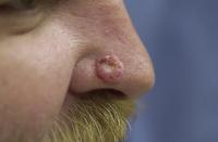 Will typically occur on light exposed areas of the body, but can be found anywhere. Eighty percent will occur on the face. Though it occurs frequently, there are few fatalities. However, basal cell cancers can grow both horizontally and vertically resulting in the destruction of any tissue they invade.
There are three primary types of Basal Cell Carcinoma: Superficial, Nodular, and Sclerosing. While these occur most commonly, there are others forms which are best diagnosed by your doctor.
The Squamous Cell Carcinoma:
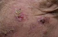 Squamous cell cancers can migrate to other parts of the body and can be potentially hazardous, however this is atypical. Differentiating between these tumors is difficult. If you have any doubt, see a dermatologist.
The three primary forms of Squamous cell carcinomas are Bowen’s Disease, Keratoacanthoma, and Infiltrating tumors. In addition, there are other forms that you should also be aware of.