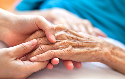
Actinic Keratoses
Actinic
keratoses are rough, red, lesions usually found on the light exposed
areas of the body particularly the face and upper extremities in
fair complexioned people which are precursors of squamous cell
carcinoma. They can occur as solitary lesions or can be found in
considerable numbers. They are the direct result of excessive sun
exposure in the patients' early childhood and young adult life.
Once they start to appear, there is a propensity for them to increase
in number even if sun exposure is significantly curtailed. The
rough spots can often be felt before actually seen. As the lesions
develop, they enlarge, the surface becomes rougher and more elevated.
A
cutaneous horn is an old actinic keratosis with a hard horny projection
overlying the lesion. The horn can be quite large. As these lesions
continue to mature, a nodule not infrequently develops under the
horn and pain or tenderness become apparent. These lesions have
become squamous cell carcinoma in situ (limited to the epidermis).
With more time, invasive squamous carcinoma develops.
Many forms of destructive therapy are available to deal with
these lesions and are extremely effective. Maintenance therapy
at regular intervals is often recommended since new lesions continue
to develop. These same patients are much more likely to develop
basal cell carcinomas as well. Detecting these cancers when they
are small, and before they exhibit tissue destruction, is certainly
in the patient's best interest. If you think you might have any
of these lesions, please see a dermatologist.
Click
here for more pictures of actinic keratoses
Alopecia Areata
Alopecia
areata is the sudden and complete loss of hair in coin shaped areas
on a hair bearing area of the skin, most commonly the scalp. It
is most common in children but can appear up to the age of forty.
It is felt to be evidence of autoimmunity but can also be influenced
by the increased pressures of everyday life (stress). An area,
once it occurs, will normally slowly enlarge for 4 to 6 weeks,
just sit there for 4-6 weeks and then gradually fill in within
the next six weeks. The older the patient, this process can be
stretched out to six months.
Unfortunately, as one area grows in, another one can fall out.
These bald spots can continue to develop resulting in significant
hair loss. Therapy is quite effective for any one spot but the
condition can be recurrent. The sooner therapy is instituted; the
more successful it's likely to be.
Click
here for more pictures of alopecia areata
Bacterial Infections
Bacterial infections of the skin are very common among skin problems.
Impetigo is often encountered. It is common in children, but no
age group is exempt. It is contagious particularly within the family.
There are two types. First, impetigo
contagiosa which presents with honey colored crusts (scabs)
and superficial erosions often on the face but can be found on
any part of the body. The second, but less common, is bullous
impetigo presenting with solitary fragile blisters which rupture
easily leaving raw, weeping, denuded areas which spread peripherally.
New blisters sprout in the surrounding area. Prompt antibiotic
therapy is rapidly curative.
Bacterial infections involving hair follicles are extremely common
particularly in shaved areas such as the beard, scalp and legs
but can occur in any hairy area. They may present as folliculitis,
superficial pustules (whitehead with a hair in the center), a furuncle green-black
necrotic plug (core) that is tenaciously embedded. Little pus is
present. When the infection is deep in the follicle at the hair
bulb level, the surrounding subcutaneous tissue is involved usually
opening in one draining sinus exuding pus. This is a boil.
When a much larger area is involved with multiple draining openings,
we refer to it as a carbuncle.
If pus is localized, it should be drained surgically and then appropriate
antibiotic therapy instituted. Antibiotic therapy without surgical
draining will often be ineffective.
Bacterial
paronychial (around the cuticle) infections are common particularly
in nail biters, nurses, and after a manicure. These are exquisitely
painful and present redness, swelling and acuminated pus under
the cuticle or lateral nail fold of a finger. It is very important
these be treated early with drainage and antibiotics. If neglected,
a felon may
develop (a deep bacterial infection in the tendon spaces of the
hand) which can prove extremely destructive with significant
morbidity and loss of function.
Most
bacterial infections are pustules, but this is more of a guideline
than a rule. The picture on the left may look like a bacterial
infection, but it is actually fire ant bites.
Click
here for more pictures of bacterial infections
Contact Dermatitis
Contact
dermatitis is certainly one of the more common skin problems dermatologists
encounter. The cause may be something the patient is allergic to
or something that is a primary irritant.
Poison ivy is a common culprit. Findings on the skin characteristically
are linear vesicular (blistering) streaks of a very pruritic (itchy)
dermatitis anywhere on the body where exposed to the plant or its
juice. Once started, it can crescendo rapidly covering extensive
surface areas with considerable to the quality of life. The sooner
therapy is instituted, the quicker the response. The problem is
recurrent with re-exposure to the plant. Effective desensitization
is not available at this time.
With other contact dermatitis, look for uncommon geometric shapes
such as straight lines, right angles, tear drops and other lesion
configurations seen as unnaturally occurring conditions. When your
suspicion is arisen, take a good history.
Click
here for more pictures of contact dermatitis
Erythema Nodosum
Erythema nodosum refers to the development of multiple painful
nodules in recurrent crops on the pretibial areas bilaterally.
Sixty percent of cases are idiopathic (without apparent cause).
In the other forty percent, the skin lesions are simply the cutaneous
manifestations of a systemic disease. What other diseases are associated
with erythema nodosum depend on the geographic area you are in.
In the Midwest, streptococcal infections predominate. In Scandinavia,
tuberculosis should be looked for. In the Philippines, leprosy
is likely. Southwest of this country, coccidiomycosis is likely
and in the Southeast, North American blastomycosis is likely. Drug
eruptions to iodides, bromides, penicillin, sulfa drugs and birth
control pills may be associated with erythema nodosum. This is
an important disease to recognize. With appropriate therapy, the
problem clears quickly.
Fungal Infections: Tineas
Fungus
infections are extremely common in clinical practice. Fungi are
plants and like all plants they like to grow in a warm, moist place.
Like flowers, there are many different fungi. In practice, fungal
infections are categorized according to the area of the body involved.
- Tinea capitis (scalp)
- Tinea corporis or ringworm (body)
- Tinea cruris or jock itch (groin)
- Tinea pedis or athletes feet (feet)
- Tinea manuum (hand)
- Tinea facei (face)
- Onychomycosis (nails)
The
key to diagnosis for all of these fungal infections is the KOH
exam. This is the ability to find the plants under the microscope
when scrapings of scale, hair or nail are soaked in KOH (potassium
Hydroxide solution). If the clinical lesion has no scaling and
you can't do a KOH, it's not a fungus infection anyway. If the
KOH is positive, the diagnosis is correct and the therapy will
work. If it's negative, therapy for a fungus infection will not
work. If a KOH is not done, the diagnosis is an educated guess.
Embarking on an expensive course of therapy without doing a KOH
exposes the patient to the risk of side effects without the likelihood
of clinical benefits. This is particular true in treating nail
disease.
Click
here for more pictures of fungal infections (tineas)
Hand Dermatitis
Hand
dermatitis is among the most common conditions dermatologists are
asked to deal with. It consists of a spectrum of diseases from
very mild annoyances with dryness and irritation, to a severe disabling
condition. Dermatitis by itself means inflammation of the skin.
This is an all-encompassing category of disease. We need some modifiers
to better classify these problems. Eczema refers to a red, denuded
and weepy dermatitis normally with considerable complaints of severe
itching. This would then be an eczematous dermatitis. Many occur
in isolated patches separated by normal appearing skin. When found
with the above, it would be called a patchy eczematous dermatitis.
Since these areas are often denuded of the epidermis, body fluid
(serum) leaks from these areas. This is a great culture media for
bacteria, which are always present on the skin.
Impetigo (superficial bacterial infection of the skin) is the
result. When combined with the above characteristic, we have an
impetiginized, patchy eczematous dermatitis.
The palms and soles have great numbers of sweat glands. With
increasing inflammation in the skin, the sweat ducts which carry
sweat to the surface of the skin is compressed like standing on
a garden hose. The sweat distends the duct below the obstruction
producing deep-seated blisters at varying depths. In its common
form, this is referred to as dyshidrosis and in its more severe
form, it is called pomphylox. The blister fluid is clear with the
composition of sweat.
There are times when the palms and soles are covered with deep-seated
pustules which occur with pustular psoriasis and its various subtypes.
Therapy for these problems is quite effective and can usually limit
disability and improve the quality of life.
Click
here for more pictures of hand dermatitis
Kaposi's Sarcoma
Kaposi's
sarcoma is the result of infection with the Human Herpes Virus
type 8. In the pre 1980's, Kaposi's was an uncommon disease of
elderly white males of a northern Mediterranean origin. Although
considered a cancer, it evolved slowly over a number of years with
low morbidity and mortality.
Over the past 25 years, latent viral infection was discovered
in a much larger segment of the population. It only became clinically
significant when the immune system was suppressed by the AIDS virus
or chemotherapy. It appears more as a tissue reaction pattern than
a true malignancy. Interestingly, wide spread disease can regress
completely when chemotherapy is discontinued and the immune system
is allowed to recover.
Today, its recognition is very helpful in identifying patients
with HIV infections. It may actually be the first clinical clue
suggesting the correct underlying diagnosis.
Click
here for more pictures of kaposi's sarcoma
Leg Ulcers
Leg
ulcers are a significant problem leading to major inconvenience,
discomfort and inability to work or function. Most leg ulcers occur
about the ankles and are secondary to varicosities (varicose veins).
This propensity runs in the family and is associated with being
over weight in the majority of cases. Leg ulcers often develop
after the age of forty in patients who spend a lot of their time
on their feet. Phlebitis (blood clots) in these legs is common.
These ulcers are called stasis dermatitis secondary to chronic
edema (fluid in the legs) and venous insufficiency. Therapy is
very effective for these ulcers. The sooner they are attended to,
the easier it is for the patient.
The second most common cause of leg ulcers is leukocytoclastic
vasculitis. This produces deep, punched out, very painful ulcers
with surrounding white scar tissue (atrophe blanche) which is pathognomonic
of this condition. These ulcers are tough to heal and are often
recurrent. You should pick the most experienced dermatologist to
handle these problems.
Arterial ulcers are the third and most difficult ulcers to manage.
They result from inflammatory vasculitis of many varied etiologies.
Embolization (blocks the flow of blood) can be caused by a blood
clot, cholesterol, tumor or arteriosclerosis (thickening and hardening
of the arterial walls). An obstructive block (total occlusion)
can lead to gangrene (death of tissue). Without sufficient blood
supply, tissue dies and the body is unable to heal these ulcers.
Peripheral vascular surgeons are usually required to deal with
these problems.
Click
here for more pictures of leg ulcers
Lichen Planus
Lichen
planus is a relatively common skin disease which often goes undiagnosed.
Usually found in middle aged adults (30 to 60), it is commonly
located on the wrists and ankles but can occur anywhere on the
body including the scalp, in the mouth or on the genitals. The
lesions are often grouped and consist of flat topped, shiny, polygonal
papules with a distinct violaceous color and a lacy whiteness over
the surface. The eruption can be extremely itchy. When the scalp
is involved, permanent scarring with hair loss can be appreciable.
When the mucous membranes of the mouth or genital areas are involved,
annular or circular lesions are found, often without symptoms.
White opalescence reticulated patterns are commonly found which
are quite diagnostic.
Several forms of the disease can be very difficult to manage.
Erosive (ulcerations) lichen planus of the mouth often requires
aggressive therapy. It can be an extremely painful condition. Hypertrophic
lichen planus consists of large, thickened, scaly plaques on the
pretibial areas (shins) which produce severe itching and without
effective therapy, can go on for twenty years or more.
Click
here for more pictures of lichen planus
Molluscum Contagiosum
Molluscum
contagiosum is a very common viral infection which is often seen
in children, tanning bed users, wrestlers and as a sexually transmitted
disease of the genital areas of young adults. Multiple lesions
are usually found and have a very typical clinical pattern. They
are well demarcated, smooth, dome-shaped, firm lesions with a central
dimple, pit or crater in its center. They can be transferred to
others by close skin to skin contact. Patients may have hundreds
of individual lesions especially in immuno-compromised patients
(patients with HIV infection, on chemotherapy, transplant patients,
etc.).
Therapy is very effective. The sooner people are treated, the
better. The lesions can spontaneously disappear but because we
cannot predicate when, how many will develop, how much it will
interfere with the quality of life or how many others will acquire
the problem from the patient, no time should be wasted in treating
them.
Click
here for more pictures of molluscum contagiosum
Paget's Disease
Paget's disease is a relatively uncommon condition usually involving
the nipple and areolar area of the breast. It is almost invariably
found in association with papillary intraductal carcinoma (breast
cancer) in the same breast. Early diagnosis and treatment is imperative
if the problem is to be treated effectively. Any breast lesion
involving the nipple should be properly evaluated by a dermatologist.
Occasionally, Paget's disease can occur in extramammary locations
usually in the groin or perirectal area, in either males or females.
These are often associated with a cancer in a nearby organ. These
lesions often go undiagnosed for a long period of time. Any skin
problem that is not resolved within 4-6 weeks could benefit from
a dermatology consult.
Click
here for more pictures of paget's disease
Perioral Dermatitis
Perioral dermatitis is a common complexion problem of young adults,
particularly women 21 to 36 which involve the areas around the
mouth and chin. It presents with small, red, itchy bumps which
come and go. It is often misdiagnosed as a dermatitis and treated
with steroid creams which may control the itching but usually makes
the problem worse. Effective therapy is readily available to control
this annoying problem Recurrences are common if therapy is discontinued
to quickly.
Click
here for more pictures of perioral dermatitis
Porokeratosis
Porokeratosis
is an uncommon tumor which occurs in two forms. Disseminated superficial
actinic porokeratosis is seen most often. It consists of extensive
flat, slightly scaling discrete lesions. They may be solitary or
literally hundreds of lesions primarily located on the light exposed
areas of the arms and legs. Adult patients who have a fair complexion
are the norm. Each of the individual lesions are surrounded by
a thickened wall of kurtosis which identify the diagnostic hallmark.
These lesions may develop into squamous cell carcinomas.
Porokeratosis of Mibelli is rarely seen but quite characteristic
clinically. It presents with a plaque normally enlarging centrically
with atrophy or thinning in the center and an elevated, warty border
around the entire lesion often referred to as the great wall of
china. Less commonly, porokeratosis type lesions can occur as a
solitary lesion on the palms and soles or in a linear pattern.
Therapy for extensive lesions can be frustrating.
Click
here for more pictures of porokeratosis
Sarcoidosis
Sarcoidosis
is a complicated disease that can mimic many others. Skin involvement
is often a central feature of this condition, although any organ
of the body can be affected. The cause is unknown to date. The
reaction pattern simulates tuberculosis and similar diseases.
The skin lesions are quite suggestive of the diagnosis. Multiple
flesh-colored or hyper- pigmented, firm papules around the central
part of the face are seen. Annular configurations about the mouth
and nose are likely to occur. Lupus pernio consisting of large
infiltrated reddish or purplish plaques on the nose or surrounding
area associated with punch out osteolytic lesions on X- ray of
the hand is quite specific. Mortality is more often determined
by the extent of pulmonary, renal and liver involvement than skin
and joint involvement.
Early recognition of this disease is essential for effective
therapy. Consultation with a dermatologist can be very helpful
in making the diagnosis as well as managing the cutaneous manifestations
of this condition.
Click
here for more pictures of sarcoidosis
Scabies
Scabies
is a common problem in society. It is caused by an itch mite acquired
through intimate contact with someone who has the disease. It is
common among school children, nursing homes, hospital personnel,
hospitalized patients and those with multiple sexual partners.
Once acquired, symptoms of a rash and severe itching develop within
thirty days. The itching becomes progressively worse particularly
at night and the rash spreads to involve the hands, between the
fingers, wrists, elbows, axillae, genitals in men, knees, ankles
and toes. This is one of the few diseases where itching of the
nipples occurs in women.
The key to diagnosis is the characteristic history and the fact
that others in the family, sexual partners, associates at work,
friends or other associates are also itching. Diagnosis is confirmed
by identifying the burrows of the active mites in the skin. It
takes an experienced clinician to find these lesions. Scabies is
often diagnosed when not actually present and not diagnosed when
it's obviously present.
Therapy is very effective. All people exposed must be treated
at the same time or recurrences are common. This is usually the
cause of treatment failure.
Click
here for more pictures of scabies
Venereal Warts
Venereal warts (genital warts) also known as condyloma acuminata
or bowenoid papulosis are transmitted from person to person during
sexual intercourse with someone who is infected with them. They
normally occur in the genital or rectal area in both men and women.
They are caused by several phenotypes of the human papilloma virus.
Infection with this virus on the cervix of women is directly correlated
with the subsequent development of cervical cancer.
It is extremely important to treat this problem as quickly as
possible before others are exposed. Men should be very considerate
of their receptive partners so they too can be evaluated. Many
forms of therapy are available. The choice is determined by the
extensiveness of the problem.
Two recent advances have occurred with this disease. One, when
women get their PAP smears for cervical cancer, they have an immunofluorescent
stain that will identify the human papilloma virus infections before
they become a problem.
The second major advance is the development of a new vaccine
that can immunize young women 8-15 to the four human papilloma
virus types that cause cervical cancer. It is currently available
and should be seriously considered for young women before their
periods start.
Click
here for more pictures of venereal warts
|